
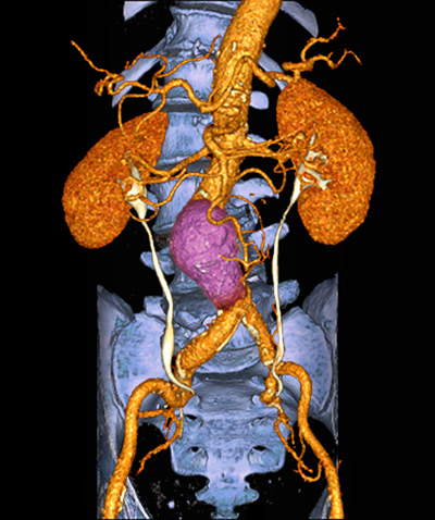
64-slice angio-CT scan of the abdomen, frontal anatomical view, showing an abdominal aortic aneurysm above the aortoiliac junction in a 71 year old male patient with high blood pressure and high cholesterol.
Abdominal pain is a common complaint that can arise from various underlying conditions affecting the organs and structures in the abdominal region. To accurately diagnose the cause of abdominal pain, medical imaging plays a crucial role in providing detailed visualization and insights into the internal structures.
Different imaging modalities, such as X-rays, CT scans, and ultrasounds, are utilized based on the suspected condition and the information required for an accurate diagnosis. In this article, we will explore the role of medical imaging in evaluating abdominal pain, focusing on X-rays, CT scans, and ultrasounds, and their specific applications.
Understanding Abdominal Pain
Abdominal pain is discomfort or pain felt in the area between the chest and the pelvis, which encompasses the abdomen. It can vary in intensity and location, and it may be associated with other symptoms such as nausea, vomiting, bloating, or changes in bowel movements. Abdominal pain can be caused by a wide range of conditions, including gastrointestinal issues, urinary tract problems, gynecological conditions, and even musculoskeletal problems.
X-rays for Abdominal Pain
X-rays are a type of medical imaging that uses small doses of radiation to produce images of the internal structures of the body. While X-rays are not the primary imaging modality for evaluating abdominal pain, they can still provide valuable information in certain situations.
Applications of Abdominal X-rays
- Obstruction. Abdominal X-rays can be used to assess for signs of bowel obstruction, which may present as dilated loops of the intestines or air-fluid levels.
- Pneumoperitoneum. In cases of a perforated viscus (such as a perforated stomach ulcer or intestinal perforation), air may leak into the abdominal cavity, causing a pneumoperitoneum. An X-ray can detect the presence of free air in the abdomen.
CT Scans for Abdominal Pain
Computed Tomography (CT) scans are powerful imaging tools that use X-rays and computer processing to create detailed cross-sectional images of the body. CT scans are highly versatile and can provide comprehensive information about the abdominal organs and structures.
Applications of Abdominal CT Scans
- Appendicitis. CT scans are often used to diagnose acute appendicitis, a condition characterized by inflammation of the appendix.
- Diverticulitis. CT scans can detect inflammation or infection in diverticula, small pouches that can form in the colon.
- Kidney Stones. CT scans are effective in visualizing kidney stones, which can cause severe abdominal pain.
- Abdominal Trauma. CT scans are valuable in assessing abdominal injuries caused by trauma, such as internal bleeding or organ damage.
- Tumors and Masses. CT scans can detect tumors or masses in the abdomen, providing detailed information about their size, location, and characteristics.
Ultrasounds for Abdominal Pain
Ultrasound imaging, also known as sonography, uses high-frequency sound waves to create real-time images of the internal organs. Ultrasounds are non-invasive, safe, and widely used for evaluating abdominal pain, especially in certain scenarios.
Applications of Abdominal Ultrasounds
- Gallstones. Ultrasounds are commonly used to detect gallstones in the gallbladder, which can cause abdominal pain and other symptoms.
- Liver and Kidney Evaluation. Ultrasounds can provide valuable information about the liver and kidneys, detecting conditions like fatty liver disease, liver cysts, or kidney stones.
- Ectopic Pregnancy. Ultrasounds are essential in diagnosing ectopic pregnancies, where a fertilized egg implants outside the uterus, often causing abdominal pain.
- Ovarian Cysts. For females experiencing abdominal pain, ultrasound can identify and assess ovarian cysts or other gynecological conditions.
Choosing the Right Imaging Modality
The choice of medical imaging modality for evaluating abdominal pain depends on various factors, including the suspected condition, the specific symptoms, the individual’s medical history, and the physician’s clinical judgment. In some cases, a combination of imaging modalities may be used to obtain a comprehensive evaluation.
- X-rays. X-rays are quick and useful for initial assessments, especially for signs of bowel obstruction or pneumoperitoneum.
- CT Scans. CT scans provide detailed images and are well-suited for diagnosing a wide range of abdominal conditions, especially when a more comprehensive evaluation is necessary.
- Ultrasounds. Ultrasounds are non-invasive and often the first choice for imaging pregnant individuals or evaluating conditions specific to certain organs like the gallbladder or uterus.
Conclusion
Abdominal pain is a common symptom that can arise from various conditions affecting the abdomen. Medical imaging plays a crucial role in diagnosing the underlying cause of abdominal pain, allowing healthcare providers to provide timely and appropriate treatment. X-rays, CT scans, and ultrasounds are essential imaging modalities, each with specific applications based on the clinical scenario.
When evaluating abdominal pain, healthcare providers carefully consider the individual’s medical history and symptoms to choose the most appropriate imaging modality to ensure an accurate diagnosis and optimal patient care.


Comments